Mouse Physiology Laboratory
Echocardiography
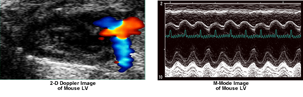
Ultrasound echocardiography provides a non-invasive measure of left ventricular (LV) dimensions and function. Mice, rats or rabbits are imaged and the EKG recorded with an Acuson Sequoia C256 instrument with a 15 MHz probe. Long axis, short axis and apical views provide two-dimensional, M-mode and spectral Doppler images at physiological heart rates. Following analysis, we can provide you with LV chamber dimensions (EDD & ESD), septal and posterior wall thicknesses (VST & PWT), ejection times, heart rates, LV masses, indices of valve function (E/A ratios) and indices of chamber function like fractional shortening, velocity of circumferential fiber shortening (Vcf), stroke volume, cardiac output and ejection fraction. Echocardiographic analyses provide a means to assess changes in the degree of ventricular hypertrophy and dysfunction over time or after other interventions such as trans-aortic constriction (see surgical section) or drug infusions.